Micro- and Nanotechnologies
Poster Session A
(1096-A) Pushing Boundaries in Cancer Research and Treatment: Innovations in the multi-Organ-on-Chip (multi-OoC) approach
Monday, February 5, 2024
12:00 PM - 1:00 PM EST
Location: Exhibit Halls AB
.png)
Abstract: In the cancer treatment, chemotherapy, pioneered by Paul Ehrlich, marked a revolutionary departure from radical surgeries. Despite a drug development, the small balance between achieving complete remission and minimizing damage to other organs remains elusive, with cancer persisting as one of a leading global cause of death. Countless promising drug investigations falter before reaching clinical stages, despite substantial investments. The primary stumbling block lies in the prevalent use of animal models, prompting the need for a paradigm shift.
In response to this challenge, the multi-Organ-on-a-Chip (OoC) approach has emerged as a transformative solution. Departing from traditional methods, this approach faithfully emulates human organ models, enabling swift drug testing, personalized therapy, and a comprehensive exploration of long-term and side effects, thereby deepening our understanding of cancer physiology.
In our in vitro research, we endeavoured to contribute to this transformative shift. Employing a multi-OoC approach, we devised a microfluidic system housing three organ models: liver, breast cancer, and heart. The objective was to construct an innovative tool for unravelling cancer physiology intricacies, evaluating anti-cancer drugs, and probing potential cardiotoxic effects.
Constructed from poly(dimethylsiloxane) (PDMS), our microsystem featured a main microchannel and three microchambers for 3D cell culture, separated by a porous polycarbonate membrane. This compartmentalization facilitated the introduction of cells into separate microchannels in the lower microchambers, while the main, upper microchannel enabled the continuous flow of nutrient medium, drugs, and intercellular communication.
To ensure physiological relevance, we developed 3D aggregates using organ-specific cell lines. The liver model comprised hepatocytes (HepG2) and hepatic stellate cells (HSC) in a 9:1 ratio. The breast cancer model integrated tumor cells (MDA-MB231) and breast fibroblasts (HMF) in a 5:1 ratio. The heart model employed cardiomyocytes (H9C2). These organ models were established by preparing and combining aggregates within a collagen type I hydrogel, subsequently introduced separately into the microsystem's lower microchambers.
Optimizing the microsystem involved different steps. The geometry of microstructures and pore distribution in membranes underwent validation using 3D measuring technology. Qualitative image analysis and extensive measurements guided the optimization of culture conditions, including medium composition and flow. Metabolic activity and viability of each organ model were assessed through AlamarBlue® and calcein-AM/propidium iodide, respectively. Techniques like CellTracker® and immunostaining were employed to analyse spatial distribution within the hydrogel.
The tested samples demonstrated consistent growth during proliferation tests, with minimal cell death observed on day 15. The optimal culture time, as derived from comprehensive data, was determined to be 12 days. CellTracker® results affirmed the accurate structure and cell ratio of the aggregates, while immunostaining confirmed the maintenance of aggregate shape throughout the 12-day culture period.
The developed microsystem presents a promising avenue, offering a viable alternative to animal testing and unlocking new dimensions for drug testing within the intricate landscape of cancer research.

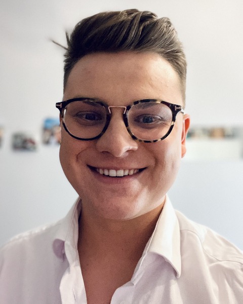
Pawel Romanczuk (he/him/his)
PhD Student
Warsaw University of Technology
Warszawa, Mazowieckie, Poland

.png)